
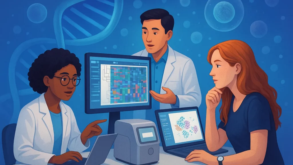

Introduction
You’ve just received a fresh single‑cell dataset. The clock is ticking, your biologist collaborators want answers by Friday, and your leadership team wants to know if there’s anything “novel” worth advancing. Do you map your cells to a reference atlas to get instant labels? Do you run de novo clustering to avoid bias? Or do you try a transfer learning model that promises both speed and nuance?
This post walks through the practical trade‑offs between atlas mapping, label transfer, and de novo analysis. We’ll keep the focus on decision‑making: how each approach performs on speed, interpretability, bias, and how well it supports discovery workflows in biotech and pharma. By the end, you’ll have a simple way to pick the right toolchain for your next project—and know when to switch gears.
What we mean by atlas mapping, label transfer, and de novo clustering
Let’s align on terms first, because teams often use them interchangeably.
Atlas mapping (sometimes called reference mapping) projects your query cells into a latent space defined by a curated cell atlas. Popular implementations include Seurat/Azimuth, Symphony, and similar “map to reference” workflows. The result is a placement of query cells alongside reference cells, often with predicted labels and confidence scores.
Label transfer is the step where labels from the reference are imputed to your query cells after alignment. It rides on atlas mapping: once cells are co‑embedded, labels move over via nearest neighbors or a classifier. It’s fast and usually accurate for common cell types and healthy tissues.
De novo analysis starts from scratch on your dataset. You build your own neighborhood graph, run community detection such as Leiden, visualize with UMAP or t‑SNE, and annotate clusters using marker genes. It’s slower than a quick map, but it avoids inheriting reference biases and is more likely to surface novel states.
Transfer learning sits between these poles. Methods like scArches and scANVI learn a shared latent space from a large reference and then adapt to your new data. Compared to straightforward label transfer, they better accommodate platform shifts and disease‑specific variation, and they can leave room for “unknown” states rather than forcing a match.
Those are the four building blocks. Most real projects mix them—often starting with a rapid map for orientation, then pivoting to de novo or transfer learning where the biology demands it.
Speed and scalability under real timelines
When deadlines are tight, start with the approach that gets you a credible answer the fastest. Reference mapping and label transfer generally win on turnaround. With a well‑chosen atlas, you can process tens of thousands of cells in minutes on a laptop and immediately hand your stakeholders a labeled UMAP. This is ideal for triage: QA the sample, confirm expected compartments, and spot glaring issues like ambient RNA or doublet‑rich clusters.
De novo analysis is slower because you must compute neighbors, run clustering, explore resolutions, and validate markers. On medium to large datasets, that’s hours rather than minutes, especially if you iterate with different normalization schemes or batch corrections.
Transfer learning lands in the middle. The cost of pretraining has already been paid by the reference authors; your job is to adapt. Fine‑tuning a scArches‑style model or applying scANVI to incorporate a partially labeled set often finishes within typical workday budgets for 50k–200k cells, especially on a single GPU. Beyond that scale—or with multi‑modal inputs—you may lean on more specialized infrastructure, but for most drug discovery datasets the compute footprint is manageable.
One more practical speed tip: choose references close to your biology. Mapping PBMCs to a blood atlas is fast and stable; mapping tumor biopsies to a healthy cross‑tissue atlas is slower and noisier. The closer the match, the fewer misalignments you’ll need to chase down.
Interpretability, bias, and labels
Speed helps you get to “first look” results, but the hard part is producing labels you can defend in a meeting with biologists, clinicians, and program leads.
Atlas mapping plus label transfer is easy to explain. You’re standing on the shoulders of a vetted reference and inheriting its controlled vocabulary. That clarity is powerful for cross‑study comparability and for downstream analyses that assume standardized labels. However, it introduces bias. If your dataset contains a new cell state, a reference‑first pipeline may confidently mislabel it as the “closest” known type. In discovery settings, that’s risky: you can miss the very signal you were tasked to find.
De novo clustering is the antidote to reference bias. It empowers you to call “unknown” clusters, characterize them with markers, and only then cross‑reference atlases as supporting evidence. It’s also the safest path in disease, development, and perturbation screens, where biology departs from healthy references. The trade‑off is interpretability cost early on. Novel clusters demand more manual work—differential expression, gene set enrichment, and targeted validation—to reach labels everyone trusts.
Transfer learning offers a compromise. Semi‑supervised models can propagate high‑confidence labels where the reference is relevant and explicitly flag out‑of‑distribution neighborhoods. This gives you coherent labeling without flattening meaningful differences. It’s often the best middle path when you suspect novelty but still need to align with a shared taxonomy.
Think of it like this: reference mapping is a shortcut through a familiar city; de novo is an exploratory hike off‑trail; transfer learning is a guided path that updates as the landscape changes. The right choice depends on how far from the city you expect to travel.
Discovery in biotech and pharma
Discovery teams care about two outcomes: not missing a therapeutic signal, and not chasing artifacts. That tension shapes your analysis strategy.
Start with a quick map to confirm sample quality and broad composition. If everything aligns and your question is routine—say, comparing T cell subset frequencies across treatments—reference mapping plus label transfer may be sufficient and auditable. You can pair it with pseudobulk differential expression and get robust, explainable results.
But if you’re profiling tumors, inflamed tissue, differentiation systems, CRISPR screens, or cell therapies, assume deviation from healthy references. Reference‑only labeling will tend to compress disease‑activated states into their nearest healthy neighbors. In these contexts, it’s wiser to treat reference mapping as a compass, not a destination. Use it to anchor obvious compartments, then switch to de novo clustering of the ambiguous fraction and test whether those groups carry distinct programs, clonotypes, or perturbation responses.
Transfer learning becomes especially useful across batches, platforms, and species. If your preclinical data lives in mouse and your clinical data in human, a transfer model trained on a joint atlas can carry over conserved programs while letting species‑specific patterns emerge. Similarly, for cross‑platform studies (e.g., 10x to full‑length protocols) or multi‑modal profiles (RNA + ATAC + proteins), transfer models frequently reduce batch headaches while preserving biological gradients—exactly what you need when go/no‑go decisions depend on subtle shifts.
A practical decision flow you can
Here’s a lightweight routine that balances speed with discovery value without relying on long checklists.
First, run a rapid atlas map to get bearings. If ≥80% of cells receive high‑confidence labels that make biological sense, bank those labels and proceed to your primary analysis. Keep an eye on the confidence distribution and the spatial coherence of predictions on the UMAP; scattered labels often hint at platform drift or batch artifacts.
Second, isolate the ambiguous slice: low‑confidence predictions, “unknown” categories, or clusters that collapse multiple tissue compartments. Re‑analyze this subset de novo. Recompute neighbors, try a small grid of resolutions, and use restrained marker testing to decide whether those groups are real states or technical composites. If they’re real, name them in plain language first (“inflammatory macrophage‑like”), then trace literature hypotheses.
Third, if ambiguity spans donors, conditions, or species, fit a transfer learning model. Provide the model with any high‑confidence labels you trust; let it learn the rest. This typically sharpens boundaries and reduces batch structure without forcing everything into the reference mold. As a bonus, you’ll get calibrated uncertainty that helps you prioritize follow‑up validation.
Finally, close the loop. Fold validated de novo clusters back into your internal reference so the next project starts one step ahead. Iteration is the real compounder of speed.
Two minimal snippets to get you started
Keep code simple when you’re making a go/no‑go choice. These examples are intentionally short; they won’t cover all your QC and batch steps but will get you oriented quickly.
Example 1: fast reference mapping and label transfer in R with Seurat/Azimuth concepts
library(Seurat)
# Query: raw counts -> SCT or log-normalize as per your SOP
qry <- CreateSeuratObject(counts = qry_counts)
qry <- NormalizeData(qry) |> FindVariableFeatures() |> ScaleData() |> RunPCA()

# Load a compatible reference object with PCA/UMAP and labels
ref <- LoadH5Seurat("path/to/reference.h5seurat")

# Anchor, map, and transfer labels
anchors <- FindTransferAnchors(reference = ref, query = qry, dims = 1:50)
qry <- MapQuery(anchorset = anchors, reference = ref, query = qry,
                refdata = list(celltype = "celltype"), reference.reduction = "pca")

DimPlot(qry, reduction = "ref.umap", group.by = "predicted.celltype")
Example 2: quick de novo clustering in Python with Scanpy
import scanpy as sc
adata = sc.read_h5ad("query.h5ad")
sc.pp.normalize_total(adata, target_sum=1e4); sc.pp.log1p(adata)
sc.pp.highly_variable_genes(adata, n_top_genes=3000, flavor="seurat_v3")
adata = adata[:, adata.var['highly_variable']]
sc.pp.scale(adata, max_value=10); sc.tl.pca(adata, svd_solver='arpack')
sc.pp.neighbors(adata, n_neighbors=20, n_pcs=50)
sc.tl.leiden(adata, resolution=0.6); sc.tl.umap(adata)
sc.pl.umap(adata, color=['leiden'])
These are starting points, not prescriptions. Your SOP for normalization, doublet detection, and batch correction should slot in before or after as appropriate.
Common failure modes and how to notice them early
Platform and chemistry mismatches are the silent killers of clean maps. When mapping outputs look speckled—labels swiss‑cheesed across UMAP space—suspect batch effects. Shifting to transfer learning or re‑embedding de novo usually resolves this faster than pushing more aggressive integration knobs.
Healthy references can over‑simplify disease. If tumor immune cells keep mapping to generic “T cell” with high confidence while your de novo analysis shows clear gradients, trust the gradients. It’s common for activation programs, exhaustion, or therapy‑induced phenotypes to fall between atlas categories.
Small atlases look deceptively authoritative. A narrow reference with few donors can make your labels seem consistent, but consistency is not correctness. Always sanity‑check with marker genes and, when available, multimodal cues like surface proteins or TCR/BCR clonotypes.
Lastly, novelty often hides in the tails. Rare clusters with modest cell counts can be genuine and actionable, especially in screens and cell therapy manufacturing lots. Before discarding them as noise, examine mitochondrial content, doublet scores, and a short panel of canonical markers. If they pass those tests and recur across donors or replicates, elevate them for validation.
Summary / Takeaways
If you need speed and standardization, start with atlas mapping and label transfer. It’s the fastest way to orient yourself, align with shared taxonomies, and deliver early results stakeholders can read.
If you need discovery and you suspect your biology deviates from healthy baselines, prioritize de novo clustering on the ambiguous slice. It preserves genuine novelty and reduces the risk of confidently mislabeling the signal you care about.
If you need robustness across platforms, donors, and species—or you want calibrated uncertainty—lean on transfer learning. It inherits the strength of references without forcing a match where it doesn’t exist.
In practice, blend them. Map first for bearings, analyze the uncertain fraction de novo, and bring in transfer learning when variation spans batches or species. Then feed your validated findings back into an internal reference so the next project moves even faster.
What dataset are you wrestling with right now—healthy PBMCs, tumors, perturbation screens, or something trickier? Share a few details about tissue, platform, and hypothesis, and I’ll suggest a tailored first‑pass plan you can run this week.
Further Reading
- Integrated analysis of multimodal single-cell data (Seurat v4, atlas mapping and label transfer) — Hao et al., Cell 2021. https://pubmed.ncbi.nlm.nih.gov/34062119/
- Efficient and precise single-cell reference atlas mapping with Symphony — Kang et al., Nature Communications 2021. https://www.nature.com/articles/s41467-021-25957-x
- Mapping single-cell data to reference atlases by transfer learning (scArches) — Lotfollahi et al., Nature Biotechnology 2022. https://www.nature.com/articles/s41587-021-01001-7
- Probabilistic harmonization and annotation with deep generative models (scANVI) — Xu, Lopez, Gayoso et al., Molecular Systems Biology 2021. https://www.embopress.org/doi/10.15252/msb.20209620
- SCANPY: large‑scale single‑cell gene expression data analysis — Wolf et al., Genome Biology 2018. https://genomebiology.biomedcentral.com/articles/10.1186/s13059-017-1382-0